Marchantia
systematic
Position of Marchantia
Kingdom- Plantae
Division- Hepaticophyta (Liverworts)
Subdivision- Hepaticae
Class- Hepaticopsida
Order- Marchantiales
Family- Marchantiaceae
Genus- Marchantia
Habitat and Distribution of Marchantia
It is one of the most common liverworts found in moist,
shady, cool areas with abundant moisture. It grows in large mats. Damp soil,
streams, springs, wet rocks are the favorable places for its growth. There are
about 65 species of Marchantia and
are found all over the world. The most common species of Himalayan region
are M. palmata, M. polymorpha and M. nepalensis.
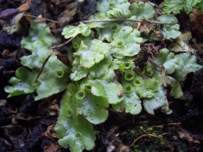
Morphology of Marchantia (Gametophyte
Phase)
<!--[if !supportLists]-->·
<!--[endif]-->Thallus is dark-green, fleshy, flat, dichotomously branched with
dorsiventral symmetry. Each thallus lobe is traversed by a central
midrib.
<!--[if !supportLists]-->·
<!--[endif]-->It also has a notch at the apex which is called as “apical notch.”
<!--[if !supportLists]-->·
<!--[endif]-->The upper surface of the thallus possesses rhomboidal or polygonal
shaped areolae. Each areola has a tiny dot in the centre and that is the air
pore that helps in aeration of the thallus.
<!--[if !supportLists]-->·
<!--[endif]-->The small cup-like structures called gemma cup are seen on the upper
surface of the thallus which is responsible for vegetative reproduction.
<!--[if !supportLists]-->·
<!--[endif]-->When the thallus attains maturity, they bear umbrella shaped structures
at the apices of certain lobes. These are called gametophores and are of two
types- antheridiophore and archegoniophore.
<!--[if !supportLists]-->·
<!--[endif]-->The antheridiophore bears antheridia and archegoniophore bears
archegonium.
<!--[if !supportLists]-->·
<!--[endif]-->From the lower surface (ventral surface) of the thallus arise the
rhizoids. These serves as an organ of anchorage and absorption. These are of
two types- smooth walled and tuberculate.
<!--[if !supportLists]-->·
<!--[endif]-->Besides the rhizoids, the ventral surface bears purplish flattered
scales. These are usually arranged in two or four rows on either side of the
thallus.
Morphology
of Marchantia. Male and Female Plant.
Anatomy of Marchantia (Internal
Structure)
The internal structure of Marchantia is
differentiated into three distinct layers:
<!--[if !supportLists]-->·
<!--[endif]-->The epidermal region
<!--[if !supportLists]-->·
<!--[endif]-->The photosynthetic region
<!--[if !supportLists]-->·
<!--[endif]-->The storage region
The epidermal region
<!--[if !supportLists]-->·
<!--[endif]-->It consists of well-defined upper and lower epidermis. The upper
epidermis forms the layer over the photosynthetic region.
<!--[if !supportLists]-->·
<!--[endif]-->It consists of single layer of thin walled cells with slightly thickened
outer walls. The epidermal cells are protective in function but they also
contain few chloroplasts. Embedded in the epidermis, there are special
barrel-shaped air pores. It facilitates gaseous exchange necessary for
photosynthesis and respiration.
The photosynthetic region
<!--[if !supportLists]-->·
<!--[endif]-->Beneath the upper epidermis is the air chambers which are arranged in
single horizontal layer. The chambers are bounded by one cell thick partitions.
From the floor of each chamber arise short, simple or branched filaments of
green cells which are known as assimilatory or photosynthetic filaments.
<!--[if !supportLists]-->·
<!--[endif]-->The photosynthetic filament contains numerous chloroplasts. It is the
principal centre of photosynthesis in the thallus. Photosynthesis is at its
maximum rate in dim light.
The storage region
<!--[if !supportLists]-->·
<!--[endif]-->Just beneath the photosynthetic region lies the storage region of the
thallus. It consists of uniform tissue made up of relatively large, colorless,
thin-walled polygonal parenchymatous cells that are compactly arranged.
<!--[if !supportLists]-->·
<!--[endif]-->They contain starch, protein grains and some of the cells contain
oil-bodies.
<!--[if !supportLists]-->·
<!--[endif]-->The lowermost layer of the storage region is composed of cells similar
to that of upper epidermis. This layer is the lower epidermis. From this layer,
rhizoids and scales arise.
Reproduction in Marchantia
Marchantia reproduces both
vegetatively and sexually.
Vegetative Reproduction in Marchantia
The methods of vegetative reproduction are as follows:
<!--[if !supportLists]-->1. <!--[endif]-->Fragmentation- The cells in
the older portions die of old age and eventually disorganize. When the death
and decay reaches dichotomy, the young lobes becomes separated. Each of these
grows into a new thallus.
<!--[if !supportLists]-->2. <!--[endif]-->Adventitious branches– In some species of
Marchantia, special adventitious branches arise from the ventral surface of the
thallus. In M.palmata, development of
adventitious branches from the stalk and disc of female gametophore has been
reported. They get detached from the parent thallus by the decay of the
connecting tissue and form new plants.
<!--[if !supportLists]-->3. <!--[endif]-->Gemma formation– Gemmae are small
bud like structures that are produced in large numbers. They are detached and
carried by wind, water and eventually grows into new individuals in new habits. In Marchantia, gemma
is a cup-shaped structure with fringed margins that is why it is known as gemma
cup and it contains many buds. Each gemma stands on a short stalk and possesses
mucilage hairs. The gemma are detached from the parent plant due to absorption
of water and carried out by water current and each gemma eventually germinates
into a new individual.
Sexual Reproduction in Marchantia
The sexual reproduction is oogamous type. It takes place only once
during the growing season in high humidity when the days are long.
Position and distribution of sex organs
The sex organs are borne on the vertical branches that are highly
specialized for this purpose. The sexual branches are apical or terminal in
position. Each upright sexual branch is called gametophore or gametangiophore.
The gametangiophore in Marchantia are
unisexual or dimorphic or heterothallic. The one bearing Antheridia is called
antheridiophore and the one bearing archegonia is called archegoniophore. The
antheridiophore and archegoniophore are borne on different thalli.
Antheridiophore- It consists of a
stalk bearing disc at the terminal region. The stalk is long and cylindrical.
The male receptacle is flattened lobed disc. Generally, it consists of eight
lobes. The antheridial chamber lies deep inside the upper surface of each lobe.
They are arranged in acropetal order. The antheridial chamber opens by a narrow
channel called ostiole.
Antheridium- The mature
antheridium is ovoid object raised on a short, multicellular stalk. The stalk
attaches to the antheridium to the floor of antheridial chamber. The body of
antheridium has a jacket layer of sterile jacket which encloses androcyte
mother cells. Each androcyte mother cell divides to form sperm cells.
Dehiscence- Presence of moisture
is necessary for the dehiscence of sperms. Water enters from the ostiole into
the antheridial chamber. The cells at the apex of this chamber absorb water and
disintegrate to form a pore. The sperms are then extruded out through the pore
and swim freely in thin film of water to reach archegonia.
Archegoniophore- It consists of a
stalk bearing disc at the terminal region. The stalk is long and cylindrical
and resembles that of antheridiophore, however the stalk is usually longer. The
female disc is an inconspicuously eight lobed object. The growing apices of
these bend downward. From the margin of the disc grows long cylindrical
processes called the rays. The archegonia are developed in acropetal order and
covered by thin layer called perichaetium.
Archegonium- The archegonium
is a flask- shaped structure that consists of a swollen portion called venter
and a slender neck.
The venter forms the swollen part. The stalk is short and helps in
attachment of archegonium to the receptacle.
Next to this, there exists a vertical row of four cells, the neck canal
cells surrounded by a layer of sterile cells forming a protective jacket.
The tip of the neck consists of four specialized large cells, known as
the cover cells. The venter also has a jacket of sterile cells which
makes up the venter wall. The venter cavity consists of two cells, the larger
one is the egg cell and is situated at the lower side and the upper one is
ventral canal cell which is smaller in size.Dehiscence- When
the archegonium reaches maturity, the ventral canal cells and the neck canal
cells degenerate to form mucilage. This mucilage imbibes water and swells which
leads to the opening up of four cover cells thus making a passage for sperms.
Fertilization in Marchantia
Fertilization takes place in presence of water. The male and female
receptacles are borne in different plants, thus it is important for both the
plants to grow together. It usually takes place by splash cup mechanism.
In this method, the sperms splashed on the grounds by the rain drops
from the male receptacle swim through the water and reach the archegonia.
However, sperms are also discharged by small insects, mites etc. In the
meantime, the neck canal cells disintegrate to form mucilage and absorb water
to swell which leads to opening of cover cells. Thus a passage is formed. The
mucilage in archegonium contains chemical substances that attract the sperms
and due to chemical interaction, they enter the venter and the most compatible
one penetrates the egg and unites to form zygote.
Sporophyte
It comprises of zygote, embryo, and sporogonium.
Zygote
<!--[if !supportLists]-->·
<!--[endif]-->It is a unicellular structure formed by the fusion of male and female
gametes and is the pioneer structure of the sporophytic phase.
<!--[if !supportLists]-->·
<!--[endif]-->It secretes a wall around it and enlarges in size.
<!--[if !supportLists]-->·
<!--[endif]-->The zygote is retained within the venter and has a diploid nucleus with
cellulose cell wall around it.
Embryo
<!--[if !supportLists]-->·
<!--[endif]-->The zygote undergoes repeated division and cell enlargement.
<!--[if !supportLists]-->·
<!--[endif]-->A spherical mass of undifferentiated cell is formed which is known as
embryo.
<!--[if !supportLists]-->·
<!--[endif]-->The venter expands as a close envelope, two cell layers thick and forms
the calyptra over the developing embryo.
Sporogonium
<!--[if !supportLists]-->·
<!--[endif]-->The sporophyte of Marchantia consists
of foot, seta and capsule.
The zygote divides by a horizontal wall at right angles to the axis of
archegonium that leads to the formation of epibasal and hypobasal regions. The
capsule is formed from epibasal region. Again, further cell division takes
place to form quadrant stage and octant stage respectively. The following
changes can be seen in the surrounding tissue as well-
<!--[if !supportLists]-->·
<!--[endif]-->The ventral cell undergoes periclinal division to form calyptra.
<!--[if !supportLists]-->·
<!--[endif]-->Perigynium eventually forms cylindrical sheath.
<!--[if !supportLists]-->·
<!--[endif]-->Perichaetium and rays further develops forming a protective covering
around whole group of archegonia.
<!--[if !supportLists]-->·
<!--[endif]-->The four epibasal cell by repeated cell division and cell
differentiation forms the capsule.
<!--[if !supportLists]-->·
<!--[endif]-->The hypobasal cells give rise to foot and seta.
<!--[if !supportLists]-->·
<!--[endif]-->The cells in the capsule region by periclinal division give rise to
different layers-
<!--[if !supportLists]-->·
<!--[endif]-->The outer layer of cells is called ampithecium and the inner one is
called endothecium.
<!--[if !supportLists]-->·
<!--[endif]-->The cells of ampithecium are undergoes division to form capsule
wall.
<!--[if !supportLists]-->·
<!--[endif]-->The cells of endothecium give rise to sporogenous tissue called archesporium.
<!--[if !supportLists]-->·
<!--[endif]-->This archesporium matures and divides to form spore mother cells.
<!--[if !supportLists]-->·
<!--[endif]-->Each spore mother cell divides meiotically to form four spores that are
arranged in tetrahedral manner and also known as spore tetrad.
<!--[if !supportLists]-->·
<!--[endif]-->However, few cells differentiate and elongate to form elaters.
<!--[if !supportLists]-->·
<!--[endif]-->In a mature spore, there are two spore coats, the outer one is exine and
the inner one is intine.
<!--[if !supportLists]-->·
<!--[endif]-->At maturity, this layer separates to release spores.
<!--[if !supportLists]-->·
<!--[endif]-->The young sporogonium is protected by three sheaths i.e. perigynium, the
calyptra and the perichaetium.
Life Cycle of Marchantia
The life cycle of Marchantia includes
two different generations, the sporophyte and the gametophyte.
Sporophytic generation is diploid and depends completely upon
gametophytic generation.
Gametophytic generation is haploid and is the dominant phase in life
cycle of Marchantia. Both the generations are
morphologically dissimilar to one another so this type of alternation of
generation is called heterologous or heteromorphic alternation of
generation.
These two individuals occur one after the other generation after
generation.
Economic Importance of Marchantia
<!--[if !supportLists]-->·
<!--[endif]-->Soil Formation and Stabilization
Marchantia plays a
critical role in soil formation by breaking down rocks and organic matter,
which contributes to the creation of new soil. It also helps in stabilizing
soil in moist environments, preventing erosion and maintaining soil structure.
<!--[if !supportLists]-->·
<!--[endif]-->Ecological Indicators
Marchantia species are
sensitive to environmental changes, making them valuable bioindicators. They
can be used to monitor ecosystem health and detect pollution levels, providing
early warnings of environmental degradation.
<!--[if !supportLists]-->·
<!--[endif]-->Medicinal Uses
Marchantia contains
compounds with antibacterial, antifungal, and anti-inflammatory properties.
These medicinal properties have been recognized in traditional herbal medicine
and are currently being explored for potential pharmaceutical applications.
Roig Y. Mesa (1945) mentioned that Marchantia polymorpha is
used to cure pulmonary tuberculosis and afflictions of the liver.
<!--[if !supportLists]-->·
<!--[endif]-->Habitat and Biodiversity
Marchantia supports
biodiversity by providing habitat and food for various microorganisms and small
invertebrates. This contribution is essential for maintaining ecological
balance and promoting nutrient cycling within their ecosystems.
<!--[if !supportLists]-->·
<!--[endif]-->Biotechnological Applications
The liverworts have played an important role at research tools in
various phases of botany such as genetics, morphology and physiology. Marchantia polymorpha is a prominent model
organism in plant biology and genetics. Its simple structure and ease of
cultivation make it ideal for studying plant development, gene function, and
responses to environmental stress.
References
<!--[if !supportLists]-->1. <!--[endif]-->Bryophyte by B.R.
Vashishta, A.K. Sinha, Adarsh Kumar (S.Chand & Company ltd)
<!--[if !supportLists]-->2. <!--[endif]-->Hait, G., Bhattacharya,
K., & Ghosh, A. K. (2012). A textbook of Botany, Volume I.
<!--[if !supportLists]-->3. <!--[endif]-->Inoue, H. 1960.
Studies in the spore germination and earlier stages of gametophyte development
in Marchantiales. Jour. Hattori Bot. Lab. 23 : 148-191.
What will i learn?
- On completion of this topic students will be able to Distinguish and identify the marchantiaene Understand the mode reproduction.
- able to understand the morphology anatomy and reproduction of algae

Write a public review